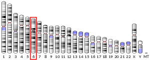
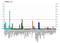
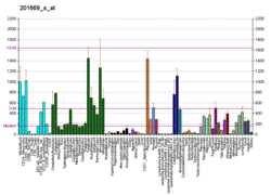
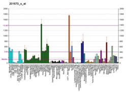
Myristoylated alanine-rich C-kinase substrate is a protein that in humans is encoded by the MARCKS gene.[3][4][5] It plays important roles in cell shape, cell motility, secretion, transmembrane transport, regulation of the cell cycle, and neural development.[6] Recently, MARCKS has been implicated in the exocytosis of a number of vesicles and granules such as mucin and chromaffin. It is also the name of a protein family, of which MARCKS is the most studied member. They are intrinsically disordered proteins, with an acidic pH, with high proportions of alanine, glycine, proline, and glutamic acid. They are membrane-bound through a lipid anchor at the N-terminus, and a polybasic domain in the middle. They are regulated by Ca2+/calmodulin and protein kinase C. In their unphosphorylated form, they bind to actin filaments, causing them to crosslink, and sequester acidic membrane phospholipids such as PIP2.
The protein encoded by this gene is a substrate for protein kinase C. It is localized to the plasma membrane and is an actin filament crosslinking protein. Phosphorylation by protein kinase C or binding to calcium-calmodulin inhibits its association with actin and with the plasma membrane, leading to its presence in the cytoplasm. The protein is thought to be involved in cell motility, phagocytosis, membrane trafficking and mitogenesis.[5]
Interactions
MARCKS has been shown to interact with TOB1[7] and with NMT2.[8]
References
- ^ a b c GRCh38: Ensembl release 89: ENSG00000277443 - Ensembl, May 2017
- ^ "Human PubMed Reference:". National Center for Biotechnology Information, U.S. National Library of Medicine.
- ^ Hartwig JH, Thelen M, Rosen A, Janmey PA, Nairn AC, Aderem A (April 1992). "MARCKS is an actin filament crosslinking protein regulated by protein kinase C and calcium-calmodulin". Nature. 356 (6370): 618–22. Bibcode:1992Natur.356..618H. doi:10.1038/356618a0. PMID 1560845. S2CID 4252140.
- ^ Blackshear PJ (January 1993). "The MARCKS family of cellular protein kinase C substrates". The Journal of Biological Chemistry. 268 (3): 1501–4. PMID 8420923.
- ^ a b "Entrez Gene: MARCKS myristoylated alanine-rich protein kinase C substrate".
- ^ Prieto D, Zolessi FR (January 2017). "Functional Diversification of the Four MARCKS Family Members in Zebrafish Neural Development". Journal of Experimental Zoology Part B: Molecular and Developmental Evolution. 328 (1–2): 119–138. doi:10.1002/jez.b.22691. PMID 27554589. S2CID 13263249.
- ^ Jin Cho S, La M, Ahn JK, Meadows GG, Joe CO (May 2001). "Tob-mediated cross-talk between MARCKS phosphorylation and ErbB-2 activation". Biochemical and Biophysical Research Communications. 283 (2): 273–7. doi:10.1006/bbrc.2001.4773. PMID 11327693.
- ^ Selvakumar P, Lakshmikuttyamma A, Sharma RK (2009). "Biochemical characterization of bovine brain myristoyl-CoA:protein N-myristoyltransferase type 2". Journal of Biomedicine & Biotechnology. 2009: 907614. doi:10.1155/2009/907614. PMC 2737134. PMID 19746168.
Further reading
- Aderem A (August 1995). "The MARCKS family of protein kinase-C substrates". Biochemical Society Transactions. 23 (3): 587–91. doi:10.1042/bst0230587. PMID 8566422.
- Herget T, Brooks SF, Broad S, Rozengurt E (October 1992). "Relationship between the major protein kinase C substrates acidic 80-kDa protein-kinase-C substrate (80K) and myristoylated alanine-rich C-kinase substrate (MARCKS). Members of a gene family or equivalent genes in different species". European Journal of Biochemistry. 209 (1): 7–14. doi:10.1111/j.1432-1033.1992.tb17255.x. PMID 1396720.
- Sakai K, Hirai M, Kudoh J, Minoshima S, Shimizu N (September 1992). "Molecular cloning and chromosomal mapping of a cDNA encoding human 80K-L protein: major substrate for protein kinase C". Genomics. 14 (1): 175–8. doi:10.1016/S0888-7543(05)80301-5. PMID 1427823.
- Harlan DM, Graff JM, Stumpo DJ, Eddy RL, Shows TB, Boyle JM, Blackshear PJ (August 1991). "The human myristoylated alanine-rich C kinase substrate (MARCKS) gene (MACS). Analysis of its gene product, promoter, and chromosomal localization". The Journal of Biological Chemistry. 266 (22): 14399–405. PMID 1860846.
- Graff JM, Stumpo DJ, Blackshear PJ (July 1989). "Characterization of the phosphorylation sites in the chicken and bovine myristoylated alanine-rich C kinase substrate protein, a prominent cellular substrate for protein kinase C". The Journal of Biological Chemistry. 264 (20): 11912–9. PMID 2473066.
- Herget T, Oehrlein SA, Pappin DJ, Rozengurt E, Parker PJ (October 1995). "The myristoylated alanine-rich C-kinase substrate (MARCKS) is sequentially phosphorylated by conventional, novel and atypical isotypes of protein kinase C". European Journal of Biochemistry. 233 (2): 448–57. doi:10.1111/j.1432-1033.1995.448_2.x. PMID 7588787.
- Taniguchi H, Manenti S, Suzuki M, Titani K (July 1994). "Myristoylated alanine-rich C kinase substrate (MARCKS), a major protein kinase C substrate, is an in vivo substrate of proline-directed protein kinase(s). A mass spectroscopic analysis of the post-translational modifications". The Journal of Biological Chemistry. 269 (28): 18299–302. PMID 8034575.
- Rao PH, Murty VV, Gaidano G, Hauptschein R, Dalla-Favera R, Chaganti RS (1994). "Subregional mapping of 8 single copy loci to chromosome 6 by fluorescence in situ hybridization". Cytogenetics and Cell Genetics. 66 (4): 272–3. doi:10.1159/000133710. PMID 8162705.
- Taniguchi H, Manenti S (May 1993). "Interaction of myristoylated alanine-rich protein kinase C substrate (MARCKS) with membrane phospholipids". The Journal of Biological Chemistry. 268 (14): 9960–3. PMID 8486722.
- Palmer RH, Schönwasser DC, Rahman D, Pappin DJ, Herget T, Parker PJ (January 1996). "PRK1 phosphorylates MARCKS at the PKC sites: serine 152, serine 156 and serine 163". FEBS Letters. 378 (3): 281–5. doi:10.1016/0014-5793(95)01454-3. PMID 8557118. S2CID 46417714.
- Swierczynski SL, Blackshear PJ (September 1996). "Myristoylation-dependent and electrostatic interactions exert independent effects on the membrane association of the myristoylated alanine-rich protein kinase C substrate protein in intact cells". The Journal of Biological Chemistry. 271 (38): 23424–30. doi:10.1074/jbc.271.38.23424. PMID 8798548.
- Spizz G, Blackshear PJ (September 1997). "Identification and characterization of cathepsin B as the cellular MARCKS cleaving enzyme". The Journal of Biological Chemistry. 272 (38): 23833–42. doi:10.1074/jbc.272.38.23833. PMID 9295331.
- Qi Q, Rajala RV, Anderson W, Jiang C, Rozwadowski K, Selvaraj G, et al. (March 2000). "Molecular cloning, genomic organization, and biochemical characterization of myristoyl-CoA:protein N-myristoyltransferase from Arabidopsis thaliana". The Journal of Biological Chemistry. 275 (13): 9673–83. doi:10.1074/jbc.275.13.9673. PMID 10734119.
- Jin Cho S, La M, Ahn JK, Meadows GG, Joe CO (May 2001). "Tob-mediated cross-talk between MARCKS phosphorylation and ErbB-2 activation". Biochemical and Biophysical Research Communications. 283 (2): 273–7. doi:10.1006/bbrc.2001.4773. PMID 11327693.
- Li Y, Martin LD, Spizz G, Adler KB (November 2001). "MARCKS protein is a key molecule regulating mucin secretion by human airway epithelial cells in vitro". The Journal of Biological Chemistry. 276 (44): 40982–90. doi:10.1074/jbc.M105614200. PMID 11533058.
- Rauch ME, Ferguson CG, Prestwich GD, Cafiso DS (April 2002). "Myristoylated alanine-rich C kinase substrate (MARCKS) sequesters spin-labeled phosphatidylinositol 4,5-bisphosphate in lipid bilayers". The Journal of Biological Chemistry. 277 (16): 14068–76. doi:10.1074/jbc.M109572200. PMID 11825894.
- Alonso S, Dietrich U, Händel C, Käs JA, Bär M (February 2011). "Oscillations in the lateral pressure of lipid monolayers induced by nonlinear chemical dynamics of the second messengers MARCKS and protein kinase C". Biophysical Journal. 100 (4): 939–47. Bibcode:2011BpJ...100..939A. doi:10.1016/j.bpj.2010.12.3702. PMC 3037601. PMID 21320438.
- Lippoldt J, Händel C, Dietrich U, Käs JA (October 2014). "Dynamic membrane structure induces temporal pattern formation". Biochimica et Biophysica Acta (BBA) - Biomembranes. 1838 (10): 2380–90. doi:10.1016/j.bbamem.2014.05.018. PMID 24866013.


