| Greater trochanter | |
|---|---|
) Upper extremity of right femur viewed from behind and above. | |
| Details | |
| Origins | vastus lateralis |
| Insertions | obturator internus, gemelli, piriformis, gluteus minimus, gluteus medius |
| Identifiers | |
| Latin | trochanter major |
| TA98 | A02.5.04.005 |
| TA2 | 1364 |
| FMA | 32852 |
| Anatomical terms of bone | |
)
The greater trochanter of the femur is a large, irregular, quadrilateral eminence and a part of the skeletal system.
It is directed lateral and medially and slightly posterior. In the adult it is about 2-4 cm lower than the femoral head.[1] Because the pelvic outlet in the female is larger than in the male, there is a greater distance between the greater trochanters in the female.
It has two surfaces and four borders. It is a traction epiphysis.[2]
Surfaces
The lateral surface, quadrilateral in form, is broad, rough, convex, and marked by a diagonal impression, which extends from the postero-superior to the antero-inferior angle, and serves for the insertion of the tendon of the gluteus medius.
Above the impression is a triangular surface, sometimes rough for part of the tendon of the same muscle, sometimes smooth for the interposition of a bursa between the tendon and the bone. Below and behind the diagonal impression is a smooth triangular surface, over which the tendon of the gluteus maximus lies, a bursa being interposed.
The medial surface, of much less extent than the lateral, presents at its base a deep depression, the trochanteric fossa (digital fossa), for the insertion of the tendon of the obturator externus, and above and in front of this an impression for the insertion of the obturator internus and superior and inferior gemellus muscles.
Borders
The superior border is free; it is thick and irregular, and marked near the center by an impression for the insertion of the piriformis.
The inferior border corresponds to the line of junction of the base of the trochanter with the lateral surface of the body; it is marked by a rough, prominent, slightly curved ridge, which gives origin to the upper part of the vastus lateralis.
The anterior border is prominent and somewhat irregular; it affords insertion at its lateral part to the gluteus minimus.
The posterior border is very prominent and appears as a free, rounded edge, which bounds the back part of the trochanteric fossa.
Additional images
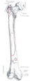
Right femur. Anterior surface.
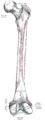
Right femur. Posterior surface.
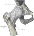
Right hip-joint from the front.
Capsule of hip-joint (distended). Posterior aspect.
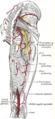
The arteries of the gluteal and posterior femoral regions.

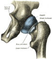
Hip joint. Lateral view.Greater trochanter.
)
)
)
See also
References
This article incorporates text in the public domain from page 244 of the 20th edition of Gray's Anatomy (1918)
- ^ Standring, Susan, editor. Gray’s Anatomy: The Anatomical Basis of Clinical Practice. Forty-First edition, Elsevier Limited, 2016, p. 1327.
- ^ Lovell, Wood W.; Winter, Robert B.; Morrissy, Raymond T.; Stuart L. Weinstein (2006). Lovell and Winter's Pediatric Orthopaedics. Lippincott Williams & Wilkins. p. 990. ISBN 978-0-7817-5358-6.
External links
- Anatomy figure: 13:01-07 at Human Anatomy Online, SUNY Downstate Medical Center
- Cross section image: pelvis/pelvis-e12-15—Plastination Laboratory at the Medical University of Vienna
- lljoints at The Anatomy Lesson by Wesley Norman (Georgetown University) (hipjointanterior, hipjointposterior)
- "Anatomy diagram: 02240.006-3". Roche Lexicon - illustrated navigator. Elsevier. Archived from the original on 2012-07-22.