)
The stratum spinosum (or spinous layer/prickle cell layer)[1] is a layer of the epidermis found between the stratum granulosum and stratum basale.[2] This layer is composed of polyhedral keratinocytes.[3][4] These are joined together with desmosomes.[3] Their spiny (Latin, spinosum) appearance is due to shrinking of the microfilaments between desmosomes that occurs when stained with H&E. Keratinization begins in the stratum spinosum,[5] although the actual keratinocytes begin in the stratum basale.[4] They have large pale-staining nuclei as they are active in synthesizing fibrilar proteins, known as cytokeratin, which build up within the cells aggregating together forming tonofibrils. The tonofibrils go on to form the desmosomes, which allow for strong connections to form between adjacent keratinocytes. The stratum spinosum also contains Langerhans cells.[6][7]
Clinical significance
Diffuse hyperplasia of the stratum spinosum is termed acanthosis.
Additional images

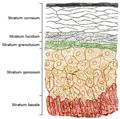
Epidermis and dermis of human skin
Section of epidermis
)
)
See also
Spinous cell
References
- ^ Rook's textbook of dermatology. Rook, Arthur., Burns, Tony, FRCP. (7th ed.). Malden, Mass.: Blackwell Science. 2004. pp. 3.7–3.8. ISBN 0-632-06429-3. OCLC 55097298.
- ^ James, William D. (William Daniel), 1950- (2005). Andrews' diseases of the skin : clinical dermatology. Berger, Timothy G., Elston, Dirk M., Odom, Richard B., 1937- (10th ed.). Philadelphia: Saunders Elsevier. p. 2. ISBN 0-7216-2921-0. OCLC 62736861.
- ^ a b Barbieri, J. S.; Wanat, K.; Seykora, J. (2014-01-01), McManus, Linda M.; Mitchell, Richard N. (eds.), "Skin: Basic Structure and Function", Pathobiology of Human Disease, San Diego: Academic Press, pp. 1134–1144, doi:10.1016/b978-0-12-386456-7.03501-2, ISBN 978-0-12-386457-4, retrieved 2020-12-04
- ^ a b McBain, A. J.; O’Neill, C. A.; Oates, A. (2016-01-01), "Skin Microbiology☆", Reference Module in Biomedical Sciences, Elsevier, doi:10.1016/b978-0-12-801238-3.99217-1, ISBN 978-0-12-801238-3, retrieved 2020-12-04
- ^ Marks, James G., Jr. (2006). Lookingbill and Marks' principles of dermatology. Miller, Jeffrey J. (Dermatologist), Lookingbill, Donald P., 1941-, Lookingbill, Donald P., 1941- (4th ed.). Philadelphia, PA: Saunders Elsevier. p. 6. ISBN 1-4160-3185-5. OCLC 70829704.
- ^ Young, Barbara (Pathologist) (2000). Wheater's functional histology : a text and colour atlas. Heath, John W., Ph. D., Stevens, Alan (Pathologist), Lowe, J. S. (James Steven), Wheater, Paul R., Burkitt, H. George. (4th ed.). Edinburgh: Churchill Livingstone. ISBN 0-443-05612-9. OCLC 43051605.
- ^ Di Meglio, Paola; Conrad, Curdin (2016-01-01), "Psoriasis, Cutaneous Lupus Erithematosus and Immunobiology of the Skin", in Ratcliffe, Michael J. H. (ed.), Encyclopedia of Immunobiology, Oxford: Academic Press, pp. 192–203, doi:10.1016/b978-0-12-374279-7.15008-8, ISBN 978-0-08-092152-5, retrieved 2020-12-04