| Trigeminal nerve nuclei | |
|---|---|
) The cranial nerve nuclei schematically represented; dorsal view. Motor nuclei in red; sensory in blue. (Trigeminal nerve nuclei are at "V".) | |
| Details | |
| Identifiers | |
| Latin | nuclei trigemini |
| MeSH | D014278 |
| NeuroNames | 2021 |
| NeuroLex ID | nifext_11 |
| FMA | 71248 |
| Anatomical terms of neuroanatomy | |
The sensory trigeminal nerve nuclei are the largest of the cranial nerve nuclei, and extend through the whole of the midbrain, pons and medulla, and into the high cervical spinal cord.
The nucleus is divided into three parts, from rostral to caudal (top to bottom in humans):
- The mesencephalic nucleus
- The chief sensory nucleus (or "pontine nucleus" or "main sensory nucleus" or "primary nucleus" or "principal nucleus")
- The spinal trigeminal nucleus
- The spinal trigeminal nucleus is further subdivided into three parts, from rostral to caudal:
- Pars Oralis (from the Pons to the Hypoglossal nucleus)
- Pars Interpolaris (from the Hypoglossal nucleus to the obex)
- Pars Caudalis (from the obex to C2)
There is also a distinct trigeminal motor nucleus that is medial to the chief sensory nucleus.
See also
Additional images
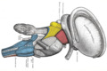
Dissection of brain-stem. Lateral view.
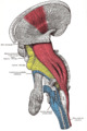
Deep dissection of brain-stem. Lateral view.
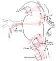
Nuclei of origin of cranial motor nerves schematically represented; lateral view.
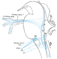
Primary terminal nuclei of the afferent (sensory) cranial nerves schematically represented; lateral view.
)
)
)
External links
- Atlas image: n2a4p5 at the University of Michigan Health System
- Washington University