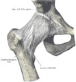
| Lesser trochanter | |
|---|---|
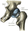
) Left hip-joint, opened by removing the floor of the acetabulum from within the pelvis. | |
) Upper extremity of right femur viewed from behind and above. | |
| Details | |
| Insertions | Psoas major, Iliacus |
| Identifiers | |
| Latin | trochanter minor |
| TA98 | A02.5.04.007 |
| TA2 | 1366 |
| FMA | 32853 |
| Anatomical terms of bone | |
The lesser trochanter of the femur is a conical eminence, which varies in size in different species.
Paleontology
The position of the lesser trochanter close to the head of the femur is one of the defining characteristics of the Prozostrodontia, which is the clade of cynodonts including mammals and their closest non-mammaliform relatives. It was erected as a node-based taxon as the least inclusive clade containing Prozostrodon brasiliensis, Tritylodon langaevus, Pachygenelus monus, and Mus musculus (the house mouse).[1]
All living mammals have a lesser trochanter, whose size, shape, and position is distinctive to their species.
Structure
In humans the lesser trochanter varies in each individual, within narrow limits. It projects from the lower and back part of the base of the femur neck.
From its apex three well-marked borders extend:
- two of these are above
- a medial continuous with the lower border of the femur neck
- a lateral with the intertrochanteric crest
- the inferior border is continuous with the middle division of the linea aspera
The summit of the lesser trochanter is rough, and gives insertion to the tendon of the psoas major muscle and the iliacus muscle.[2]
Clinical significance
The lesser trochanter can be involved in an avulsion fracture.[3]
Additional images
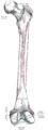
Right femur. Posterior surface.
Right hip-joint from the front.
Capsule of hip-joint (distended). Posterior aspect.

Hip joint. Lateral view. Lesser trochanter

Muscles of Thigh. Anterior views.
)
)
)
See also
References
This article incorporates text in the public domain from page 245 of the 20th edition of Gray's Anatomy (1918)
- ^ Liu, J.; Olsen, P. (2010). "The Phylogenetic Relationships of Eucynodontia (Amniota: Synapsida)". Journal of Mammalian Evolution. 17 (3): 151. doi:10.1007/s10914-010-9136-8.
- ^ Federle, Michael P.; Rosado-de-Christenson, Melissa L.; Raman, Siva P.; Carter, Brett W., eds. (2017-01-01), "Female Pelvic Floor", Imaging Anatomy: Chest, Abdomen, Pelvis (Second Edition), Elsevier, pp. 1050–1077, ISBN 978-0-323-47781-9, retrieved 2021-01-22
- ^ Khoury JG, Brandser EA, Found EM, Buckwalter JA (1998). "Non-traumatic lesser trochanter avulsion: a report of three cases". Iowa Orthop J. 18: 150–4. PMC 2378165. PMID 9807723.
External links
- Anatomy figure: 13:01-11 at Human Anatomy Online, SUNY Downstate Medical Center
- lljoints at The Anatomy Lesson by Wesley Norman (Georgetown University) (hipjointanterior, hipjointposterior)