| Lacrimal sac | |
|---|---|
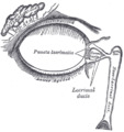
) The lacrimal apparatusis shown through dissection on the left side. | |
) The lacrimal sac has been opened showing internal organization as well as the naso-lacrymal duct. | |
| Details | |
| Artery | angular artery |
| Identifiers | |
| Latin | saccus lacrimalis |
| TA98 | A15.2.07.068 |
| TA2 | 6857 |
| FMA | 20289 |
| Anatomical terminology | |
The lacrimal sac or lachrymal sac[1] is the upper dilated end of the nasolacrimal duct,[2] and is lodged in a deep groove formed by the lacrimal bone and frontal process of the maxilla. It connects the lacrimal canaliculi, which drain tears from the eye's surface, and the nasolacrimal duct, which conveys this fluid into the nasal cavity.[3] Lacrimal sac occlusion leads to dacryocystitis. [4]
Structure
It is oval in form and measures from 12 to 15 mm. in length; its upper end is closed and rounded; its lower is continued into the nasolacrimal duct.
Its superficial surface is covered by a fibrous expansion derived from the medial palpebral ligament, and its deep surface is crossed by the lacrimal part of the orbicularis oculi, which is attached to the crest on the lacrimal bone.
Histology
Like the nasolacrimal duct, the sac is lined by stratified columnar epithelium with mucus-secreting goblet cells, with surrounding connective tissue. The Lacrimal Sac also drains the eye of debris and microbes.
Function
It serves as a reservoir for overflow of tears, in which the lacrimal sac pumps inward and outward driven by the orbicularis muscle during blinking.
Imaging
The lacrimal sac can be imaged by dacrocystography, in which radiocontrast is injected, followed by X-ray imaging.
Additional images
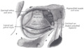
Medial wall of left orbit.
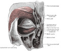
Left orbicularis oculi, seen from behind.
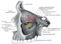
The lacrimal apparatus. Right side. (Lacrimal sac visible at upper right.)
The tarsi and their ligaments. Right eye; front view. (Lacrimal sac visible at middle right.)
)
)
)
)
See also
References
This article incorporates text in the public domain from page 1028 of the 20th edition of Gray's Anatomy (1918)
- ^ Dr. Johannes Sobotta (1909). Sobotta's Atlas and Textbook of Human Anatomy. Philadelphia.
- ^ "Endoscopic Dacryocystorhinostomy". ScienceDirect. 2019-01-01. pp. 143–148.e1. doi:10.1016/B978-0-323-47664-5.00017-1. Retrieved 2020-05-08.
Anatomy: The lacrimal sac extends approximately 10 mm above the axilla of the middle turbinate.
- ^ Remington, Lee Ann (2012). "Ocular Adnexa and Lacrimal System". Clinical Anatomy and Physiology of the Visual System. Elsevier. pp. 159–181. doi:10.1016/b978-1-4377-1926-0.10009-8. ISBN 978-1-4377-1926-0.
The lacrimal sac lies within a fossa in the anterior portion of the medial orbital wall. This fossa is formed by the frontal process of the maxillary bone and the lacrimal bone. The sac is surrounded by fascia, continuous with the periorbita, which runs from the anterior to the posterior lacrimal crests.
- ^ Beare, Nicholas A.V.; Bastawrous, Andrew (2014). "Ophthalmology in the Tropics and Sub-tropics". Manson's Tropical Infectious Diseases. Elsevier. pp. 952–994.e1. doi:10.1016/b978-0-7020-5101-2.00068-6. ISBN 978-0-7020-5101-2.
Tears drain through the canaliculi, lacrimal sac and naso-lacrimal duct into the nose. Blockage of the naso-lacrimal duct prevents drainage of the lacrimal sac, which may lead to infection, causing a painful swelling at the side of the nose below the medial canthus. This may present as a chronically watery, discharging eye, or as an acutely inflamed abscess. This should be treated with oral broad-spectrum antibiotics. The problem may recur unless drainage into the nose is re-established.