| Dorsal column nuclei | |
|---|---|
| Identifiers | |
| NeuroLex ID | nlx_153860 |
| Anatomical terms of neuroanatomy | |
| Dorsal column nuclei | |
|---|---|
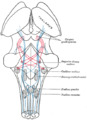
) Present at the junction between the spinal cord and medulla oblongata, the dorsal column nuclei consist of paired gracile, and cuneate nuclei (labels 6 and 7, respectively). | |
| Details | |
| System | Somatosensory system |
| Identifiers | |
| NeuroLex ID | nlx_153860 |
| Anatomical terminology | |
In neuroanatomy, the dorsal column nuclei are a pair of nuclei in the dorsal columns in the brainstem.[1] The name refers collectively to the cuneate nucleus and gracile nucleus, which are present at the junction between the spinal cord and the medulla oblongata. Both nuclei contain second-order neurons of the dorsal column-medial lemniscus pathway, which carries fine touch and proprioceptive information from the body to the brain. Each nucleus has an associated nerve tract, the gracile fasciculus and the cuneate fasciculus.
Nuclei and tracts
The dorsal column nuclei also include their tracts, or fasciculi.
Gracile nucleus
The gracile nucleus is medial to the cuneate nucleus; its neurons receive afferent input from dorsal root ganglia sensory neurons subserving the lower trunk and limbs. The gracile nucleus and gracile fasciculus carry epicritic, kinesthetic, and conscious proprioceptive information from the lower part of the body (below the level of T6 in the spinal cord). Because of the large population of neurons in the gracile nucleus they give rise to a raised area called the gracile tubercle on the posterior side of the closed medulla at the floor of the fourth ventricle.
Cuneate nucleus
The counterpart to the gracile nucleus and fasciculus is the cuneate nucleus and cuneate fasciculus, which carries the same type of information, but from the upper body (above T6, except the face and ear which is carried by the principal sensory nucleus of trigeminal nerve). The cuneate nucleus is wedge-shaped and located in the closed part of the medulla. It lies lateral to the gracile nucleus and medial to the spinal trigeminal nucleus in the medulla. The large number of neurons found there give rise to the cuneate tubercle seen on viewing the posterior aspect of the medulla on the side of the brainstem.
Neurons of the dorsal column nuclei send axons that form the internal arcuate fibers, crossing over at the sensory decussation to form the medial lemniscus, ultimately synapsing with third-order neurons of the thalamus.
Additional images
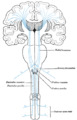
Scheme showing the course of the fibers of the lemniscus; medial lemniscus in blue, lateral in red.
The sensory tract.

Fourth ventricle. Posterior view. Deep dissection.
)
)
)
References
- ^ Standring, Susan (2016). Gray's anatomy: the anatomical basis of clinical practice (41 ed.). Elsevier Limited. pp. 309–330. ISBN 978-0-7020-5230-9.