| Femoral head | |
|---|---|
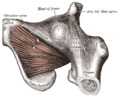
) Upper extremity of right femur viewed from behind and above. | |
| Details | |
| Identifiers | |
| Latin | Caput femoris |
| MeSH | D005270 |
| TA98 | A02.5.04.002 |
| TA2 | 1361 |
| FMA | 32851 |
| Anatomical terms of bone | |
The femoral head (femur head or head of the femur) is the highest part of the thigh bone (femur). It is supported by the femoral neck.
Structure
The head is globular and forms rather more than a hemisphere, is directed upward, medialward, and a little forward, the greater part of its convexity being above and in front.
The femoral head's surface is smooth. It is coated with cartilage in the fresh state, except over an ovoid depression, the fovea capitis, which is situated a little below and behind the center of the femoral head, and gives attachment to the ligament of head of femur.
The diameter of the femoral head is generally larger in men than in women.
Fovea capitis
The fovea capitis is a small, concave, depression within the head of the femur that serves as an attachment point for the ligamentum teres (Saladin). It is slightly ovoid in shape and is oriented "superior-to-posteroinferior. (Cerezal)" This orientation is said to be favorable for the tensed fibers of the ligamentum teres. The fovea capitis is located "slightly posterior and inferior to the center of the articular surface of the femoral head (Cerezal)" Furthermore, unlike the head of the femur, the fovea capitis lacks any hyaline cartilage. The fovea capitis is said to contain vascular canals in two-thirds of individuals, but "their contribution to femoral head vascularity varies. (Cerezal)"
Clinical significance
If there is a fracture of the neck of the femur, the blood supply through the ligament becomes crucial. In orthopedic surgery, the head of the femur is important because it can undergo avascular necrosis and consequent osteochondritis dissecans. The femoral head is removed in total hip replacement surgery.
Additional images
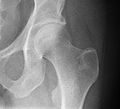
Radiograph of a healthy human hip joint
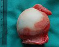
Gross pathology specimen of the head of the femur with some synovium attached at the bottom and the ligament attached at the top. Ruler in centimeters at left side.
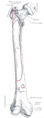
Right femur. Anterior surface.
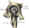
Left hip-joint, opened by removing the floor of the acetabulum from within the pelvis.
The Obturator externus.

Hip joint. Lateral view. Femur head

Hip joint. Lateral view. Femur head
)
)
)
)
)
)
References
This article incorporates text in the public domain from page 243 of the 20th edition of Gray's Anatomy (1918)
Cerezal, Luis. "Anatomy, Biomechanics, Imaging, and Management of Ligamentum Teres Injuries." RadioGraphica. RSNA, Oct. 2010. Web. Nov. 2015.
Saladin, Kenneth S. Anatomy & Physiology: The Unity of Form and Function. 6th ed. New York, NY: McGraw-Hill, 2007. 300-302. Print.
External links
- Cross section image: pembody/body15a—Plastination Laboratory at the Medical University of Vienna